Writer: Kim Burkhardt, RT, R, MR, Northwest Radiology Network
Magnetic resonance imaging (MRI) is a noninvasive medical test that helps physicians diagnose and treat medical conditions. MRI uses a powerful magnetic field, radio frequency pulses and a computer to produce detailed pictures of organs, soft tissues, bone and virtually all other internal body structures. MRI does not use ionizing radiation (X-rays).
 How Does MRI Work?
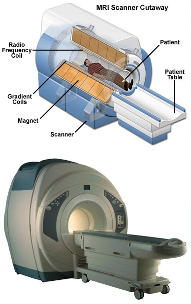
To obtain the MRI image, a patient lies down on a table inside a large magnet. Sometimes, contrast agents may be given to a patient intravenously before or during the MRI to enhance the image, clarifying certain anatomic structures or certain pathologies.
Incorporating an advanced technology, MRI produces images of the anatomy without the use of radiation, unlike in X-ray and CT scanning. MRI images are formed by computer processing signals emitted by body tissue. These signals are generated using a safe magnetic field in combination with radio waves of a specific frequency, similar to what is used in home and car radios.
MRI is a safe, painless and noninvasive exam. There are no preparations required for this exam. Some patients even fall asleep during their exam. Used for all parts of the body, MRI results in no known side or after effects.
MRI can help provide a quick and accurate diagnosis for your physician. In some situations, this can reduce the need for exploratory surgery and other diagnostic procedures which might have associated risks.
The procedure is effective in the clinical evaluation of the following conditions: brain disorders, traumatic injuries, eye abnormalities, spine diseases, tumor detection, liver and other abdominal diseases, knee and shoulder injuries, musculoskeletal disorders, facial/neck abnormalities, infection, cardiac malformations and blood flow/vessel disorders.
Northwest Radiology Network offers MRI as well as many other imaging services!